Characterisation of Synthetic Diamond Crystals
Post Date: 16 May 2011 Viewed: 1419
In the quest for perfect synthetic single crystal diamonds the novel technique of µm-scale spatially-resolved high-resolution X-ray diffractometry was employed. Why are high quality synthetic diamonds so valuable for X-ray optics applications and why is the improvement of their crystalline quality so crucial? Firstly, diamond has the best thermal properties of all materials known to man, which is of paramount importance for high heat-load optics [1]. Secondly, the applications of diamond crystals for synchrotron X-ray optics are numerous; ranging from single-deflection X-ray monochromators, double-crystal Bragg and Laue X-ray monochromators and phase plates. Moreover, with the advent of the so-called fourth-generation sources (FELs), one can expect a growing demand for high-quality diamond crystals as X-ray optical elements, since diamond seems to be one of the very few materials that will survive the extreme power density of a free electron laser X-ray beam.
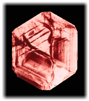
Unfortunately natural diamonds are defect-rich by nature and each natural diamond is unique. In contrast, the best presently available synthetic diamonds have a typical average mosaic spread of one to two arcseconds and can be locally perfect. The principal problem is the non-uniform distribution of defects, which causes inhomogeneous beam intensities for X-ray optical applications. The main defects are growth bands and dislocations. Stacking faults, inclusions and cleavage traces leaving strains are also observable. These features are visible in the white beam topograph of a (111)oriented, 1.2 mm thick crystal shown in Figure 109. Such defects continue to limit the range of applications of diamond. To understand better the relationship between the growth parameters, the distribution and content of nitrogen (the major impurity) and the crystalline perfection double-crystal X-ray diffractometry with a microscopic spatial resolution was applied.

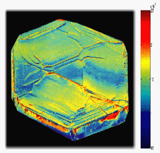
The experiment was performed at the BM5 beamline at an energy of 10 keV. Double-crystal rocking curves of a (220)-oriented perfect silicon crystal and (111)-oriented diamond crystal in almost non-dispersive geometry were recorded with a FReLoN (Fast-Readout-Low-Noise) 2D-CCD camera developed at the ESRF. Rocking curves were then attributed to each pixel so that in one angular scan the rocking curve parameters such as width, peak reflectivity and peak position could be mapped across the crystal surface with a resolution of 10 micrometres. Because the camera was fixed at a distance of 0.5 m, the scans were so-called w-scans and represented lattice tilts rather than lattice strains or d-spacing variations. The width of the rocking curves (FWHM) is presented in Figure 110 as a contour map. An unprecedented tilt resolution even smaller than the Darwin width could be achieved and maps of the d-spacing distribution could be obtained by appropriate binning of the detector pixels, without moving the camera.

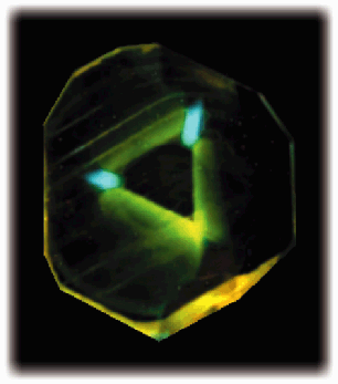
Superimposing the micro-defect structure determined by X-ray diffraction on X-ray excited optical luminescence (XEOL) images (Figure 111), a clear correlation with nitrogen impurities and defects could be demonstrated. It was confirmed that concentration variations were related to lattice imperfections, and that tilts were greater than lattice parameter variations.

This new technique enables not only a considerable enhancement of spatial resolution but also a substantial time gain of five orders of magnitude with respect to traditional rocking curve mapping [2]. Serving as a high-resolution tool for defect control and limitation in synthetic diamond crystals and to guide the future engineering developments, it will have an important impact on the synthesis of high quality diamonds.


